Key Features and Advantages of DIARM™ DM
A Mammography Dedicated Workstation
- Automatic Display Protocols
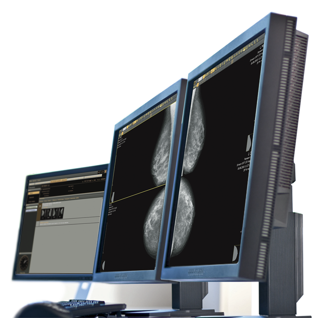
Automatic display sequencing of mammography images for screening and diagnostic studies in accordance with standard protocols. The radiologist does not need to spend time on manual placement or correcting the orientation of images. This significantly reduces the time needed for the examination of images and review summary. This is important in exam rooms with high patient traffic. As a result, the analysis of mammograms is more convenient for radiologists and increases the likelihood of detection of abnormalities during early stages.
- Tomosynthesis Module
The Tomosynthesis module displays Digital Breast Tomosynthesis exams using industry-recognized standards. The doctor can scroll through images manually or in a Cine mode. In order to remember a specific slice, the radiologist can bookmark it. The module also allows for changing the reconstruction slice thickness and the intensity projection visualization method (MIP, MinIP, MeanIP).
- Zoom and position corrections are applied to the images so that mammogram pairs are located symmetrically on the monitors, thus placing the paired areas on the images adjacent to each other.
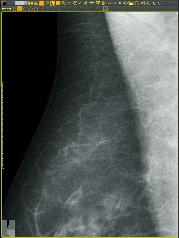
- The Pixel View tool provides a simple way to see every pixel of the image which allows a better focus on specific regions of interest.
- The Image Navigator shows the breast area that is currently displayed, helping the radiologist to identify and recall suspect locations.
- Mammography-grade 5MP Monitors
The workstation is equipped with two specialized 5 megapixel medical displays. These are advanced medical imaging monitors that can display high-resolution mammograms. In most cases, there will not be a need to scale images in order to analyze the smallest abnormalities. In addition to high-resolution, the monitors also have outstanding brightness and contrast, as well as increased life span for continuous operation without loss of performance.
f
Easy and Convenient Use
- Recall and preview historical patient studies.
- Display new and historical images simultaneously for comparison.
- Easy and fast access to Visualization Modes and Tools.
- Simple and effective real-time image filters make it possible to highlight details or suppress noise, emphasizing any potential areas of concern.


Comprehensive Image Manipulation and Workflow Optimization
- Tools for analyzing digital images including zoom, image placement on the screen and subtle brightness/contrast adjustments are all accessible through the click of a button. All features are easily accessible.
- A wide range of measuring tools help carry out numerical analysis of abnormalities: measuring the length of a segment, size of an area, and angles. All measurements can be applied to the mammogram as annotations and sent to the printer.
- User customizable report templates are embedded in the workstation.
- The fully automated workflow efficiently enables the radiologist to describe exam conclusions, make prints, and prepare a CD that includes DICOM images. A convenient and easy to use CD preparation program for viewing on standard computers is also included.

DIARM™ DM Mammography Workstation Hardware Specifications
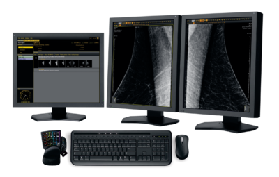
- Modern, high-performance medical workstation
- Medical-Grade Monitor, 5MP (two are included)
- High resolution, 22″ LCD color monitor
- Dedicated Multifunction Keypad
- Keyboard
- Mouse

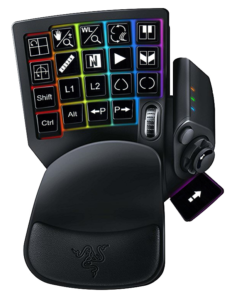
Dedicated Mammo Keypad
- A dedicated multifunction keypad is provided for fast execution of the most commonly used functions
- Facilitates the one button image review workflow process
- Simple and easy to use
- Promotes efficiency and decreases total image review time


